

Dermoid Sinus
Dermoid Sinuses are narrow tube-like structures, which are derived from a skin defect. They penetrate from the skin surface to varying depths downward into the muscles and towards the spinal cord. They are situated in the midline of the neck and croup, which is in front and behind the area occupied by the ridge. There may be several sinuses in any one puppy.
This is the only known congenital defect that occurs regularly in the breed. (Congenital meaning that the defect is formed before birth). When considered as a defect in the dog family as a whole, Dermoid Sinuses occur only very rarely in dogs, other than Ridgebacks or Crossbred Ridgebacks. It must therefore be obvious that it is an inherited defect which has become widespread in the "blood lines" of the breed as a result of the early selective breeding of the original stock from which the Ridgebacks of today have been produced.
The incidence of the defect throughout the breed is Australia is not known, as the recording of the numbers of Dermoid Sinus affected pups in litters has not been done on a scale large enough to enable a statistical analysis to be carried out. Many breeders in the past did not even record dermoid puppies produced as they were euthanized on detection of the sinus. Over the last decade or so attitude to Dermoid has changed and many breeders will have an operation performed on the puppy at 8-9 weeks with the pup subsequently sold/homed as a pet.

Areas marked X indicate the sites at which
dermoid sinuses may develop.
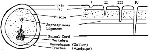
Formation of Dermoid Sinus
The sinus takes the form of a tube opening at the skin surface and running a varying depth toward the spine. The lining grows hair and has sebaceous glands - dark hair may project through the external opening. Dermoid Sinuses occur when small areas of attachment between the outer layer of cells (the skin) and the tubular structure (later to become the brain and the spinal cord) remain. In the puppy this defective separation of the embryological tissues is present as a thin tubular attachment extending from the skin of the midline of the top surface of the dog to the deeper tissues below, and as deep as the spinal cord in some cases. The depth to which this tubular skin defect penetrates is the criterion used for the classification of four types of Dermoid Sinuses.
Cross section through a dog's neck
TYPE I Penetrates below skin surface, its fatty tissue
overlying the neck muscles.
TYPE II Penetrates into the muscles of the neck.
TYPE III Penetrates to supraspinous ligament,
running over the top of the vertebrae.
TYPE IV Penetrates to the spinal cord between the
vertebrae.
.png)
Feeling for presence of Dermoid Sinus by sliding longitudinal fold of skin between index finger and thumb.
Photos courtesy of Ms Anke Terbruggen
Diagnosis of Dermoid Sinus
In the puppy, Dermoid Sinus can be detected by gently raising the skin in a longitudinal fold along the top midline in the area in which Dermoid Sinuses are known to occur (i.e. in front of and behind the ridge). If the skin fold is raised with one hand and the skin allowed to slip back and forward between the thumb and forefinger of the other hand the presence of the Sinus can be felt as a thin cord-like structure or fine thread between the two layers of skin. Raising the skin fold in this way tenses the tissues and a Dermoid Sinus will be pulled tautly between its skin attachment in the top midline and its attachment in the muscles below. Care must be taken however, to differentiate between a sinus and fine muscle strands that may be felt in the neck and shoulder region. If there is doubt, raise the skin in front of and behind the suspect area and the sinus will appear as a depressed area. Raising the skin clearly shows a depressed area and the sinus is plainly visible.
The diagnosis can be confirmed by shaving the hair from the skin over the point at which the Dermoid Sinus is attached. A small pore like opening in the skin from which a small tuft of hair protrudes is usually seen. This is the opening of the Dermoid Sinus on the skin surface. The darkened spot caused by hair protruding from the sinus is easily seen once the area has been clipped. The older the puppy, the thicker the Sinus will be and the more easily it may be recognised.
Significance of Dermoid Sinus
The detrimental effects of Dermoid Sinus are not just concerned with the fact that a visible anatomical defect is apparent in affected animals, but rather the complications which can arise as a result of a Dermoid Sinus becoming infected with bacteria. The effect on the animal varies directly in proportion to the depth of penetration i.e. ranging from boil like eruptions at skin level to severe spinal pain and paralysis. A Dermoid Sinus is a progressive condition and is not self-limiting.
The narrow tube of skin that descends below the skin surface is lined with all the normal skin structures and of special significance are hair, sweat, and oil glands. The thin central cavity that runs down the Dermoid Sinus, becomes filled in time with hair, skin oil and skin scales. The contents usually become an ideal medium where bacteria that are normally present on the skin, may grow. They gain access to the material through the small pore-like opening at the point of attachment of the Dermoid Sinus on the skin surface.
Accumulated skin secretion undergoes a process of putrefaction, and the skin barrier of the Sinus walls breaks down and bacteria invade the tissues deep below the skin surface. This usually results in the formation of an abscess that eventually ruptures to the outside and drains as a chronically discharging purulent wound. Extensive surgical and medical treatment may be necessary to clear up such a complication and, in some cases, septic Dermoid sinus may be unresponsive to treatment.
If a Dermoid Sinus is recognised in a dog before it becomes septic, it can be removed surgically, with a good chance that no further complications will occur. In most cases however, owners of animals are not aware of the presence of a Dermoid Sinus and shortly thereafter sepsis almost always sets in. Subsequently, the owners are obliged to obtain veterinary treatment to resolve the distressing complications. This may be costly to the dog owner although responsible breeders will bear some of the surgical / medical costs.
Responsible breeders check for sinus as soon as possible after birth and continue checking again and again usually also having other experienced breeders check as well, until the pups go to new home with information on Dermoid sinus provided to new owners to ensure that the condition is recognised if a sinus has been missed.
For new owners it is advisable to be ensure that vaccination injections in Ridgebacks puppies are carried out away from the midline to prevent any reaction at the vaccination site being mistaken for a sinus and, more importantly that an undetected sinus cannot be attributed to faulty injection technique.
References:
"Dermoid sinus in the Rhodesian Ridgeback (A Review by a Veterinarian)" published by South African Rhodesian Ridgeback Club.
"Guide to the Rhodesian Ridgeback" published by The Rhodesian Ridgeback Club of Great Britain.